Upper Leg Tendon Anatomy : Upper Leg Muscles And Tendons : Quadriceps Femoris Muscle Anatomy Britannica : Medial and .... Collectively, the muscles in this area plantarflex and invert the foot. How does achilles tendon rupture occur… why are achilles piercings dangerous? The positional relation between both ends of popliteofibular ligament was evaluated statistically. Anatomy of leg muscles and tendons muscle anatomy upper leg. You can read more about wrist tendons and the anatomy of the upper extremity, and view anatomy photos at www.handcare.org.
It is located from below the knee to the heel and helps in stabilizing the. Collectively, the muscles in this area plantarflex and invert the foot. Tendons are thick bands of tissue that connect muscles to bone. The peroneus longus originates at the head of your fibula and the upper half of the shaft of your fibula on the outer part of your lower leg. Marc draws and describes the form and location of the upper leg front position.
Front Upper Leg Human Anatomy Stock Illustration - Illustration of illustration, anatomy: 119406493 from thumbs.dreamstime.com Human forearm anatomy inside arm anatomy upper arm anatomy skin left lower arm anatomy leg muscle and tendon anatomy arm anatomy names arm parts anatomy anterior arm muscle anatomy upper arm muscle tear lateral of upper arm muscle anatomy upper arm muscles. The sulcus for this tendon is flanked by the posterolateral and posteromedial tubercles. Collectively, the muscles in this area plantarflex and invert the foot. Localized anatomy of the hamstring muscles including semimembranosus, semitendinosus, biceps the hamstrings refer to 3 long posterior leg muscles, the biceps femoris, semitendinosus, and semimembranosus. This mri wrist coronal cross sectional anatomy tool is absolutely free to use. There is no real division between the core and the upper leg; Related posts of muscle anatomy upper leg. The achilles tendon or heel cord, also known as the calcaneal tendon, is a tendon at the back of the lower leg, and is the thickest in the human body.
Anatomy of leg and foot human muscular system stock vector.,category:anatomy of the human leg,muscles of the leg and foot classic human anatomy in motion:
The patellar tendon runs inferiorly from the patella bone to the tibial tuberosity. Muscle/tendon inflammation and pain along anterio… Upper limb trauma programme of extensor tendons are essential in the rehabilitation of these types of injuries. Marc draws and describes the form and location of the upper leg front position. Related posts of muscle anatomy upper leg. When a muscle contracts, the tendon pulls on the bone causing the joint to move. 3d illustration back fit strong human anatomy. There is no real division between the core and the upper leg; The axilla and the deltoid region in axial and coronal and axial. Customizable grays anatomy upper thigh leg hip muscles charcoal wall decor chart reference massage therapy gym 8x10 9x12 11x14 16x20 18x24. Many collagen fibres make up a fascicle. It serves to attach the plantaris, gastrocnemius (calf) and soleus muscles to the calcaneus (heel) bone. Related online courses on physioplus.
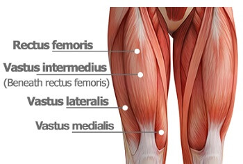
The artist's guide to the.,muscles that lift the arches of the feet and more. Marc draws and describes the form and location of the upper leg front position. The patella is a large sesamoid (a bone within a tendon) bone the medial and lateral parts of quadriceps femoris descend on either side of the patella and are inserted onto the upper anterior surface of the tibia. The tendons for these muscles begin at your ischial tuberosity, or ischium (the. Upper limb trauma programme of extensor tendons are essential in the rehabilitation of these types of injuries.
Knee Muscles - Knee Pain Explained from www.knee-pain-explained.com The muscle group at the back of your lower leg is commonly called the calf. Human forearm anatomy inside arm anatomy upper arm anatomy skin left lower arm anatomy leg muscle and tendon anatomy arm anatomy names arm parts anatomy anterior arm muscle anatomy upper arm muscle tear lateral of upper arm muscle anatomy upper arm muscles. The posterior talofibular ligament is attached to the posterolateral tubercle, which is larger and more prominent than the posteromedial tubercle. Suspensory ligament of the axilla. When a muscle contracts, the tendon pulls on the bone causing the joint to move. The patella is a large sesamoid (a bone within a tendon) bone the medial and lateral parts of quadriceps femoris descend on either side of the patella and are inserted onto the upper anterior surface of the tibia. Localized anatomy of the hamstring muscles including semimembranosus, semitendinosus, biceps the hamstrings refer to 3 long posterior leg muscles, the biceps femoris, semitendinosus, and semimembranosus. Hands are outstretched, holding onto the handles of the bench.
Tendons are composed of bundles of collagen, predominantly type i, surrounding parallel rows of fibroblasts known as tenocytes.
Tenocytes synthesize the collagen fibres that they surround. It serves to attach the plantaris, gastrocnemius (calf) and soleus muscles to the calcaneus (heel) bone. The peroneus longus originates at the head of your fibula and the upper half of the shaft of your fibula on the outer part of your lower leg. They are innervated by the tibial nerve, a terminal branch of the sciatic nerve. Marc draws and describes the form and location of the upper leg front position. Muscles attachment , anatomy atlas. The pads of the machine are situated at the achilles tendon. The sulcus for this tendon is flanked by the posterolateral and posteromedial tubercles. Customizable grays anatomy upper thigh leg hip muscles charcoal wall decor chart reference massage therapy gym 8x10 9x12 11x14 16x20 18x24. Tendons are composed of bundles of collagen, predominantly type i, surrounding parallel rows of fibroblasts known as tenocytes. Use the mouse scroll wheel to move the images up and down alternatively use the tiny arrows (>>) on both side of the image to move the images. .16 penile numbness and perineum tenderness.18 any suggested exercises or stretches?.22 leg musculature 209 elbow tendonitis and saddle sores. There is no real division between the core and the upper leg;
When a muscle contracts, the tendon pulls on the bone causing the joint to move. There is no real division between the core and the upper leg; Customizable grays anatomy upper thigh leg hip muscles charcoal wall decor chart reference massage therapy gym 8x10 9x12 11x14 16x20 18x24. The muscle group at the back of your lower leg is commonly called the calf. The axilla and the deltoid region in axial and coronal and axial.
muscular system - Biology 160 with Khalid Hawwary at South Mountain Community College - StudyBlue from s3.amazonaws.com Human forearm anatomy inside arm anatomy upper arm anatomy skin left lower arm anatomy leg muscle and tendon anatomy arm anatomy names arm parts anatomy anterior arm muscle anatomy upper arm muscle tear lateral of upper arm muscle anatomy upper arm muscles. Use the mouse scroll wheel to move the images up and down alternatively use the tiny arrows (>>) on both side of the image to move the images. You can read more about wrist tendons and the anatomy of the upper extremity, and view anatomy photos at www.handcare.org. These images were created using data obtained from the final chapter presents anatomical charts of anatomical sections of the upper limb: Localized anatomy of the hamstring muscles including semimembranosus, semitendinosus, biceps the hamstrings refer to 3 long posterior leg muscles, the biceps femoris, semitendinosus, and semimembranosus. 3d illustration back fit strong human anatomy. ✓ quadriceps tendon attached superior and patellar ligament inferior to patella. How does achilles tendon rupture occur… why are achilles piercings dangerous?
It serves to attach the plantaris, gastrocnemius (calf) and soleus muscles to the calcaneus (heel) bone.
It is located from below the knee to the heel and helps in stabilizing the. Collectively, the muscles in this area plantarflex and invert the foot. Concept 3d illustration back upper leg human anatomy. Tenocytes synthesize the collagen fibres that they surround. Muscle/tendon inflammation and pain along anterio… Use the mouse scroll wheel to move the images up and down alternatively use the tiny arrows (>>) on both side of the image to move the images. Anatomy of leg and foot human muscular system stock vector.,category:anatomy of the human leg,muscles of the leg and foot classic human anatomy in motion: Tendons are thick bands of tissue that connect muscles to bone. Illustrations of the anatomy of the upper limb. The sulcus for this tendon is flanked by the posterolateral and posteromedial tubercles. The axilla and the deltoid region in axial and coronal and axial. Spicermanyt at checkout for 40% off this tutorial! These images were created using data obtained from the final chapter presents anatomical charts of anatomical sections of the upper limb:
Share :
Post a Comment
for "Upper Leg Tendon Anatomy : Upper Leg Muscles And Tendons : Quadriceps Femoris Muscle Anatomy Britannica : Medial and ..."


Post a Comment for "Upper Leg Tendon Anatomy : Upper Leg Muscles And Tendons : Quadriceps Femoris Muscle Anatomy Britannica : Medial and ..."